MUSCLES
INTRODUCTION
- Study of muscles known as Myology.
- Myology also known as Sarcology.
- Development of muscles : –
- Except muscles of Iris & cilliary body all muscles of body develop from mesoderm.
- Muscle of Iris, cilleary body & myoepithelial cell of sweat gland develop form Ectoderm.
- Conductivity & contractility are the two main characteristics of muscles.
Three types of muscles are found in the body.
- (i) Voluntary or skeletal muscles.
- (ii) Involuntary or smooth muscles.
- (iii) Cardiac muscles.
FORCE GENERATING PROTEIN
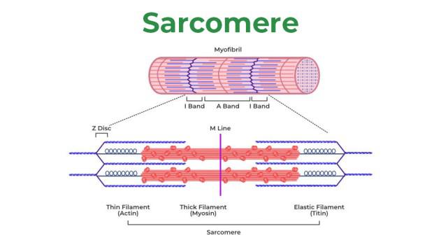
- Actin – The Actin or thin filament is a double helix made up of protein molecule called as. G-Actin (Globular actin).
- Many G-actin combined to form a filament like structure, which is called as filamentous-actin.
- G-actin contain an active site where myosin head is attached.
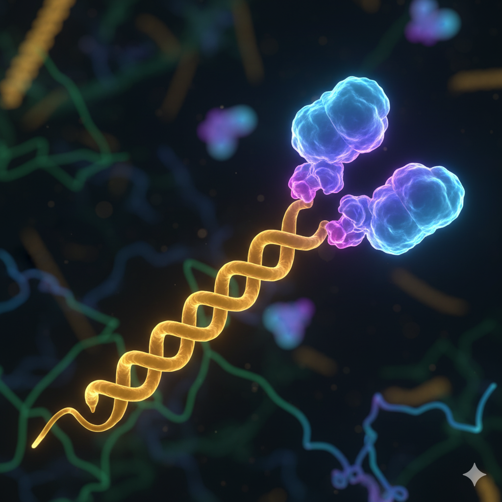
- Myosin – Each myosin molecule consists of a tail & head.
- Tail is made up of two chains intertwined with each other like double helix.
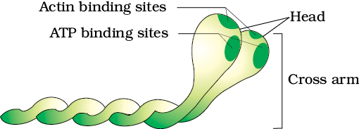
- The myosin head has sites for attachments with (i) The actin filament (ii) ATP molecules.
- Each myosin (thick) filament is also a polymerised protein.
- Many monomeric proteins called Meromyosins constitute one thick filament.
- Each meromyosin has two important parts, a globular head with a short arm and a tail.
- The former being called the heavy meromyosin (HMM) and the latter, the light meromyosin (LMM).
- The HMM component, i.e.: the head and short arm projects outwards at regular filament and is known as cross arm.
- The globular head is an active ATPase enzyme and has binding sites for ATP active sites for actin.
REGULATING PROTEIN
- Tropomyosin – It is one type of contractile protein.
- In the relaxed state of the muscle situated in such a way, that the active sites remain covered by the Tropomyosin & attached at the terminal end of actin.
- Troponin – It is one type of protein which attached with one of ends of the tropomyosin molecules.
Troponin is made up of three subunit.
- (a) Troponin I (Inhibitory site)
- (b) Troponin T (Tropomyosin site)
- (c) Troponin C (Ca+2 binding site)
STRUCTURAL PROTEIN
- Actinin – It is one type of protein which found in Z-line.
SLIDING FILAMENT THEORY
- This theory is given by A.F. HUXLEY, H.E. HUXLEY & HANSEN.
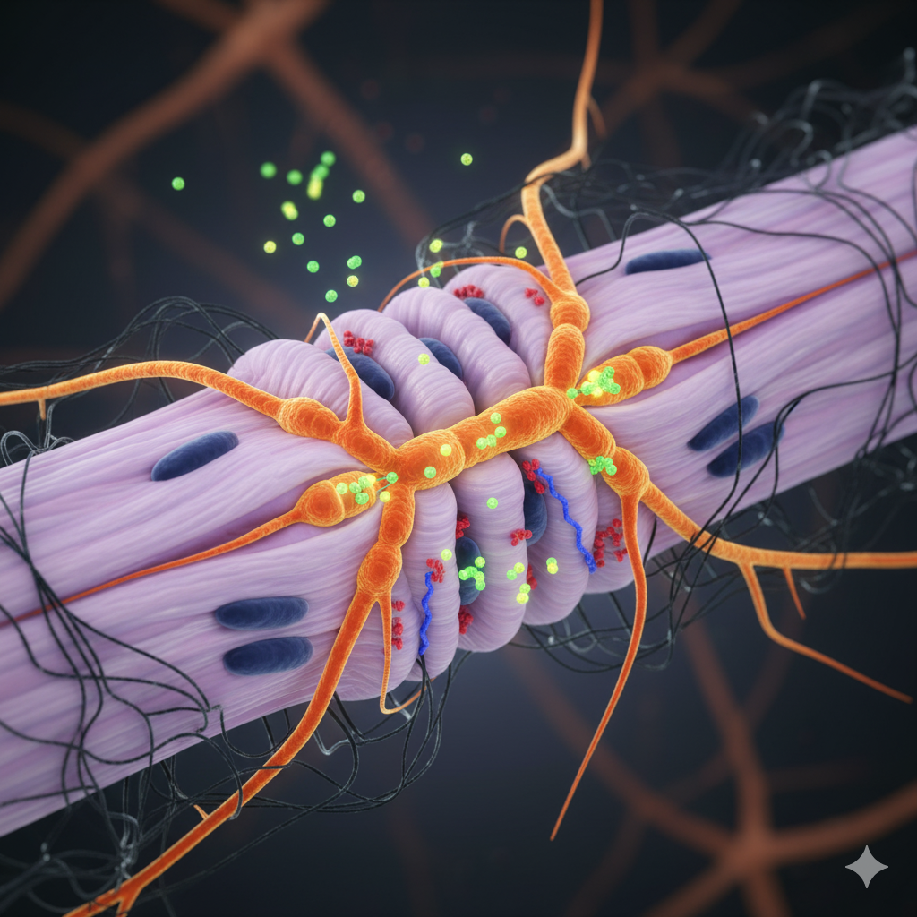
- The junction of Nerve & muscle is called as neuromuscular junction.
- Terminal branches of Axon of motor nerve is embedded into sarcolemma.
- Its bulb like structure is called as motor end plate.
- Sarcolemma invaginates inside & form a fimbriated structure which is called synaptic gutter or subneural.
- cleft.
- The cell membrane of the bulbous terminals is called as the pre junctional membrane.
- The cell membrane of muscle fibre which invaginates is called post junctional membrane.
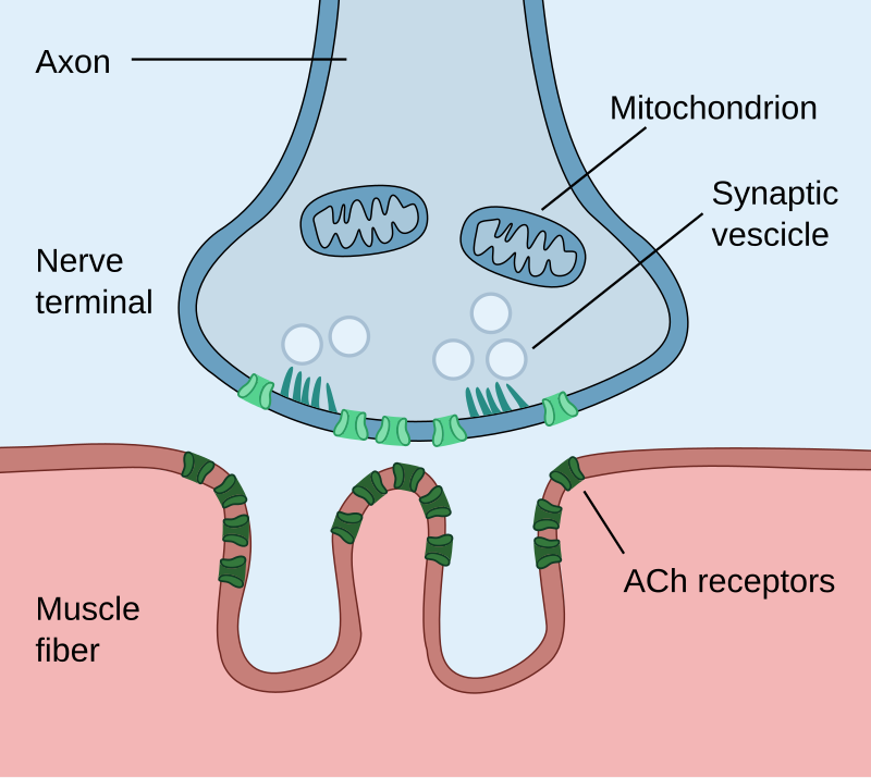
- In motor end plate large number of vesicles & mitochondria are present.
- Each vesicle contains Ach in high concentration.
- In post junctional membrane, Ach receptor are presented.
- (Ach = Acetylcholine, it is a neurotransmitter chemical).
Neuromuscular Transmission
- When motor nerve fibre stimulated it develops an Action potential. (Resting potential 50 to 100 mV).
- AP reaches in the neuromuscular junction & goes to bulbous expansion of the nerve terminal.
- Than it increases permeability of Ca++ in the Pre junctional membrane.
- Ca++ enter from E.C.F. in to the cytosol of motor end plate by penetrating the prejunctional membrane.
- Ca++ ions causes bursting of the vesicles & releases the Ach.
- These Ach now cross the prejunctional membrane.
- Ach reaches via subneural cleft the post junctional membrane.
- Ach attach the Ach receptor also called as End plate receptor.
- End plate receptor stimulate & develop end plate potential by opening of Na+–K+ channels in post synaptic membrane.
- When end plate potential sufficiently higher than A.P. develop on sarcolemma & myofibril.
- Sarcolemma invaginates inside & form transverse & longuituidnal tubules which are also called as. T-tubul and L-tubule.
- T-tubules are parallel to Z-line wherease L-tubule is perpendicular to the Z-line.
- T-Tubules communicates with ECF.
- T & L system of tubules together called as endoplasmic reticulum.
- L-Tubules dilated on both side of T-Tubules this dilated part called cisterns.
- A.P. proceeds along the sarcolemma & A.P. contact with T-Tubules.
- A.P. further proceeds via T-tubules & enter with in muscle fibre.
- This AP is now called as T-tubule potential.
- T-tubule potential come in close contact of L-tubules at region of the Triads (T + L-tubules).
- L-tubules are a very rich source & store house of Ca++ ion in higher concentration.
- The process results in the release of Ca++ ion in large amount.
- Released Ca++ ion combine with troponin C.
- In Relaxed state tropomyosin covers the active site of actin.
- When troponin-C combines with Ca++ ion some physiochemical changes occur in tropomyosin.
- Tropomyosin move away from active site of actin.
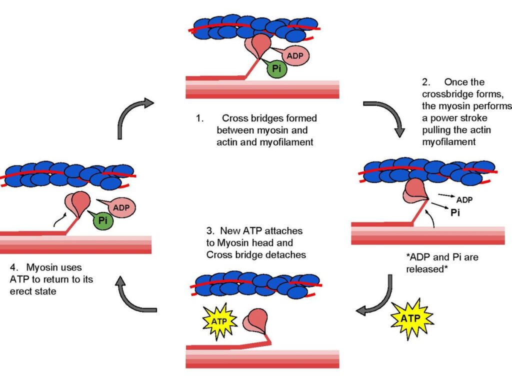
- Myosin have strong tendency to bind the actin molecule & Actomyosin complex is formed.
- Myosin head attach on active site of actin with the help of cross bridges.
- The myosin head twists in the groove of the active site of actin-F.
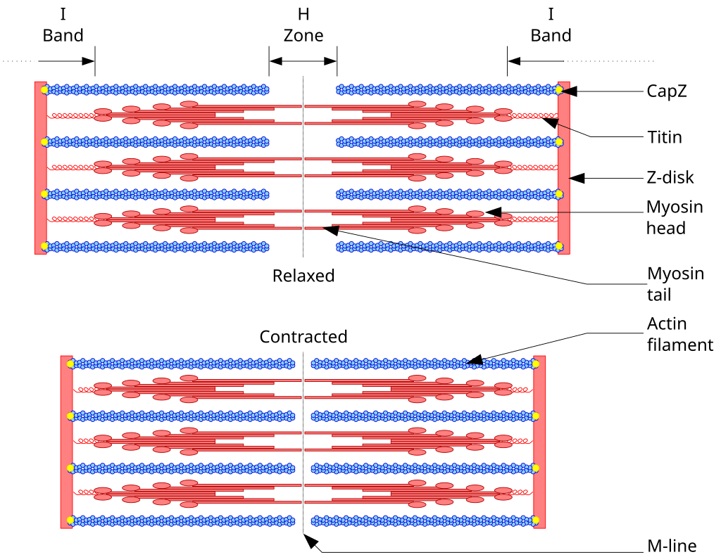
- This causes movement of actin toward H-zone.
- Contraction is caused by overlapping of actin filament over myosin – sliding filament hypothesis.
- All the cross bridges move simultaneously in one direction.
- The actin filaments move vigorously towards H-zone.
- When cross bridge disrupted than myosin molecule detached & reattach the new active site of actin.
- After muscle contraction H-Zone disappears.
- Length of sarcomere & I-band decreases by 20%.
- The length of A-band remains unchanged.
- All process are reversible; at the time of relaxation Ca++ are goes into L-tubules.
Role of ATP:-
- The Rotational movement of myosin head with in the groove.
- (Deattachment of myosin head form the actin.
Chemical reaction in Muscles: –
- ATP + H2O ¾¾¾¾ ®¾ kinaseCreatine ADP + P1 + Energy (For contractile muscle)
- Creatine phosphate + ADP ¾¾ ®¾ Creatine + ATP (Muscle contraction )
- Gycogen ¾¾¾¾ ®¾ Glyocysis Lactic acid + Energy
- 80% Lactic acid + Water ¾¾¾ ®¾ ATP Glycogen (Liver cell)
- 20% Lactic acid + Oxygen ¾¾ ®¾ CO2 + H2O + ATP (Liver cell)
- Creatine + ATP ¾¾¾ ®¾ Creatine phosphate + ADP (Resting Muscle)
PROPERTIES OF MUSCLES
Terminology
- Origin – Fixed end of muscle (Proximal end).
Insertion – Distal end of muscle which is attach to bone (Movable end).
- Excitability – Muscles responds to stimuli which can be nervous, chemical, electrical & thermal mechanical.
- Conductivity – Stimulus acting in one region of muscle fibres propagated to all parts within no time.
- Contractility – On being stimulated the muscle fibres contract & shorten followed by Relaxation.
- Threshold stimulus – Intensity of stimulus below the threshold value does not produces contraction in.
- muscle fibres is called Sub threshold Stimulus.
- A stimulus stronger than threshold one is called suprathreshold stimulus.
- All or none law – Response of muscle fibre is maximum whether the stimulus has threshold value or suprathreshold value.
- Response is absent when intensity is subthreshold (Below threshold value).
- Muscle twitch – It is single isolated contraction of Muscle fibres due to single stimulus. Muscle curve or kymograph indicates three phases.
- Period of latent excitation (Latent period).
- Latent period is the interval between the application of appropriate stimulus & initiation of contraction.
- It is 0.01 sec. in skeletal muscle.
- It is 3 sec. in smooth muscle.
- Contraction phase – Duration for which muscle remain contracted state.
- It is 0.04 sec. in skeletal muscle.
- It is 20 sec. in smooth muscle.
(c) Relaxation phase – Interval for contracted muscle to regain its original/Relaxed state.
- It is 0.05 sec. in skeletal muscle.
- It is 23 sec. in smooth muscle.
- Paralysis – Supply of motor nerve impulse completely cut off.
- Function of muscle contraction is stopped.
- Shivering – Involuntary contraction of muscles to make body warm.
- Muscle tension – force produced during contraction of muscle is known as muscle tension.
- Isometric – Contraction occur when a muscle is stimulated adequately but is prevented to shorten. e.g. applying too heavy load against the muscle so that the muscle but cannot lift the at all ext.
- Isotonic – When muscles is stimulated adequately & is allowed to shorten.
- Then the contraction is called Isotonic.
- Some external work is done.
- Technically called a load is lifted.
- Antagonistic muscles – They are pair of muscles which causes opposite movement at the same site.
- When one muscle is contracting, the other is relaxes & viceversa.
- e.g. – Biceps (flexor) & Triceps of arms (extensor).
- Motor unit – Groups of mucles fibres supplied by single motor neuron.
- It is a functional unit of muscles.
- All the muscle fibres of motor unit contract & relax simultaneously.
- Cori cyles – Lactic acid accumulated in muscles during sustained contraction.
- Formed lactic acid transported in blood as blood lactate to liver where is changes into liver glycogen.
- Liver glycogen is changed in to glucose.
- Speed of – Skeletal muscle = 0.1 sec. per contraction per cycle.
- Cardiac muscle = 0.8 sec. per contraction per cycle.
- Smooth muscle = 46 sec. per contraction per cycle.
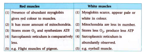
- Red muscle fibres | White muscle fibres
- Fatigue : Due to sustained contraction initially muscle give beneficial effects of contraction (warm ups).
- After warm ups, ATP is exhausted & muscle is as state of permanent contraction & no relaxation.
- This permanent state occurs because no fresh ATP available for detachment of actomysosin complex.
- Fatigue is caused B/o :
- Accumulation of lactic acids.
- Consumption of stores glycogen, ATP, CTP (Creatinine phosphate).
In fatigue:
- (i) Increase latent period and phase of relaxation.
- (ii) decrease height of contraction.
- Rigor Mortis
- After death fresh supply of ATP. become impossible.
- Due to non availability of ATP/C.P. deattachment of myosin from actin cannot take place.
- This results in permanent state of contraction of muscle.
- This phenomenon is called rigor mortis.
- This condition helps fixation of the hour of death.
- E.D.T.A (Ethylane diamine tetra acetic acid) injected inside muscle combined with Ca+ and stops contraction.
- Muscle and nerve exitability is reduced by K+.
- During muscle contraction chemical energy changed into mechanical energy.
- Over stretching of tendon is called sprain.
MUSCLE TYPES ON BASIS OF MOVEMENTS
- Flexor = Fore arm move in upward direction (Bend).
- Bending of part over one another.
- Eg. biceps brachii.
- Extensor – Fore arm move in downward direction.
- Straighting of bending part.
- Eg. Triceps.
- Adductor – Toward body axis. Towards the body.
- Lattissimus dorsi brings the arms towards body.
- Abductor-upper & lower limb move away from body axis.
- Abductor moves away from the body (midline).
- Eg. deltoids.
- Pronator – Palm state in down. Rotate downward.
- Eg. pronater teres.
- Supinator – Palm state in upward.
- Rotate upward.
- Eg. brachioradialis.
- Dilatation – Diameter increases, widening of Iris (radial muscle of iris).
- Constrictor – Diameter decreases, Closing an aperture.
- Eg. sphincter ani closes anus.
- Depressor – Lower Jaw move in downward direction.
- Lowering part.
- Eg. depressor mandibularis.
- Elevator–Lower Jaw move in downward direction. (Note: This line repeats the ‘depressor’ movement description but identifies the term as Elevator)
- Raising the part.
- Eg. Massetar.
- Median Rotation – Upper & lower limb rotate in inward direction.
- Lateral Rotation – Outward direction rotation.
- Inversion when sole of foot turn toward body axis.
- Eversion – Away from body axis.
- Aryeiglotticus muscle is called Hilton muscle.
- Gastrocenemius muscle present in shank.
- Sartorius the longest muscle of body.
- Gluteus maximus (Buttock muscles) – Largest muscle of body.
- Stapedius – smallest muscle of body.
- In Human beings 639 muscle are found.
- 634 muscle are paired and 5 muscle are unpaired.
- 400 muscles are striated.
- Most of muscles are found in back region.
- Number of back muscles are 180.
- Jaw muscles are strongest.
- Longest smooth muscles is present in present in uterus of pregnant lady.
Practice Questions
- Assertion (A): Muscle contraction involves shortening of muscle fibers as force is generated.
Reason (R): Sliding filament mechanism explains that actin and myosin filaments slide past each other to shorten sarcomeres.
Options: (a) Both A and R are true and R is the correct explanation of A. (b) Both A and R are true but R is not the correct explanation of A. (c) A is true but R is false. (d) A is false but R is true.
Answer: (a) - Assertion (A): Skeletal muscle fibers are multinucleate due to fusion of myoblasts.
Reason (R): Myoblasts are mononucleated precursor cells that fuse during development.
Answer: (a) - Assertion (A): Sarcomere is the functional unit of striated muscle.
Reason (R): Z-lines mark the boundary of each sarcomere and sliding between filaments changes sarcomere length.
Answer: (a) - Assertion (A): Motor end plate is the specialized area of muscle fiber sarcolemma.
Reason (R): Acetylcholine released from motor neuron binds to receptors on the motor end plate to initiate contraction.
Answer: (a) - Assertion (A): T tubules transmit action potentials into the interior of muscle fibers.
Reason (R): T tubules are invaginations of the sarcolemma that are closely associated with sarcoplasmic reticulum.
Answer: (a) - Assertion (A): Sarcoplasmic reticulum stores calcium ions in resting muscle.
Reason (R): Release of calcium from SR into the cytosol triggers interaction of actin and myosin.
Answer: (a) - Assertion (A): ATP binding to myosin head causes detachment from actin.
Reason (R): Hydrolysis of ATP on myosin drives conformational changes for the power stroke.
Answer: (a) - Assertion (A): Creatine phosphate is an immediate reservoir for ATP regeneration.
Reason (R): Creatine kinase catalyzes transfer of phosphate from creatine phosphate to ADP to form ATP.
Answer: (a) - Assertion (A): Oxidative phosphorylation in mitochondria provides sustained ATP during prolonged exercise.
Reason (R): Mitochondria are abundant in slow-twitch fibers that rely on aerobic respiration.
Answer: (a) - Assertion (A): Anaerobic glycolysis yields ATP rapidly but produces lactate.
Reason (R): Fast-twitch fibers rely on glycolytic pathways for short bursts of high-intensity activity.
Answer: (a) - Which of the following statements correctly describe the energy metabolism during muscle contraction?
(i) ATP hydrolysis by myosin provides immediate energy for power stroke.
(ii) Phosphocreatine acts as a rapid ATP buffer for a few seconds.
(iii) Oxidative phosphorylation in mitochondria provides long-term ATP supply.
(iv) Glycolysis requires oxygen and produces the most ATP.
(a) (i), (ii), (iii)
(b) (ii), (iii)
(c) (i), (ii), (iv)
(d) All of these
Answer: (b) - Which of the following statements about sarcomere organization are true?
(i) The A-band remains constant in length during contraction.
(ii) The I-band shortens as actin slides inward.
(iii) The H-zone disappears at full contraction.
(iv) The Z-lines move farther apart during contraction.
(a) (i), (ii), (iii)
(b) (ii), (iii)
(c) (i), (iii), (iv)
(d) (ii), (iv)
Answer: (a) - Which of the following statements are correct regarding calcium’s role in contraction?
(i) Calcium binds to troponin and removes tropomyosin blockage.
(ii) Calcium activates myosin ATPase directly.
(iii) Calcium release from SR is triggered by T-tubule depolarization.
(iv) Calcium remains permanently in the cytoplasm after contraction.
(a) (i), (iii)
(b) (ii), (iii), (iv)
(c) (i), (ii)
(d) (iii), (iv)
Answer: (a) - Which of the following statements describe events leading to muscle relaxation?
(i) Calcium is pumped back into SR using ATP.
(ii) Troponin returns to its inhibitory configuration.
(iii) Acetylcholine breakdown stops further stimulation.
(iv) Actin–myosin cross-bridges remain permanently attached.
(a) (i), (ii), (iii)
(b) (ii), (iv)
(c) (i), (iv)
(d) All of these
Answer: (a) - Which of the following statements correctly describe neuromuscular junction physiology?
(i) Synaptic vesicles release acetylcholine into the cleft upon nerve impulse.
(ii) Acetylcholine binds receptors on the motor end plate to cause depolarization.
(iii) Calcium influx into presynaptic terminal triggers transmitter release.
(iv) Acetylcholinesterase synthesizes acetylcholine.
(a) (i), (ii), (iii)
(b) (ii), (iii), (iv)
(c) (i), (iv)
(d) All of these
Answer: (a) - Which of the following statements are incorrect regarding fast-twitch fibers?
(i) They rely mainly on anaerobic glycolysis.
(ii) They fatigue more quickly than slow-twitch fibers.
(iii) They contain abundant mitochondria and myoglobin.
(iv) They are adapted for endurance activity.
(a) (iii), (iv)
(b) (i), (ii)
(c) (ii), (iii)
(d) All of these
Answer: (a) - Which of the following statements correctly describe excitation–contraction coupling?
(i) Action potential travels through T-tubules.
(ii) Dihydropyridine receptors interact with ryanodine channels.
(iii) Calcium release triggers actin–myosin interaction.
(iv) The system is independent of membrane depolarization.
(a) (i), (ii), (iii)
(b) (ii), (iii), (iv)
(c) (i), (iii)
(d) All of these
Answer: (a) - Which of the following statements are correct about muscle fatigue?
(i) It is caused by accumulation of lactic acid.
(ii) ATP levels remain constant throughout.
(iii) Fatigue results from reduced calcium sensitivity.
(iv) Neural factors contribute to reduced excitation.
(a) (i), (iii), (iv)
(b) (ii), (iv)
(c) (i), (ii)
(d) (i), (ii), (iii)
Answer: (a) - Which of the following statements about muscle energy systems are true?
(i) ATP provides energy for only a few seconds of maximal work.
(ii) Creatine phosphate rapidly restores ATP levels.
(iii) Glycolysis supports intermediate activity duration.
(iv) Oxidative phosphorylation is dominant during short bursts.
(a) (i), (ii), (iii)
(b) (ii), (iv)
(c) (i), (iii)
(d) All of these
Answer: (a) - Which of the following statements are correct about muscle fiber ultrastructure?
(i) Myofibrils are cylindrical contractile elements arranged parallel inside fibers.
(ii) Sarcomeres shorten during contraction without filament length change.
(iii) T-tubules transmit electrical signals to deeper regions.
(iv) H-zone expands during contraction.
(a) (i), (ii), (iii)
(b) (ii), (iv)
(c) (i), (iii)
(d) All of these
Answer: (a) - The contraction of skeletal muscle is initiated by a nerve impulse that reaches the neuromuscular junction. This impulse causes release of acetylcholine, which binds to receptors on the sarcolemma. What is the immediate result of this binding?
(a) Opening of sodium channels causing depolarization of the sarcolemma
(b) Release of calcium from mitochondria
(c) Activation of ATPase in actin filaments
(d) Direct stimulation of myosin head rotation
Answer: (a) - During skeletal muscle contraction, the I-band shortens while the A-band remains constant because:
(a) Thick filaments slide past thin filaments without changing length
(b) Actin filaments contract while myosin remains fixed
(c) The Z-lines disintegrate and move apart
(d) The number of sarcomeres decreases
Answer: (a) - Calcium ions play a vital role in initiating muscle contraction. Their primary function is to:
(a) Bind to troponin and shift tropomyosin from actin binding sites
(b) Attach to myosin heads to provide energy for movement
(c) Act as a neurotransmitter to stimulate motor neurons
(d) Trigger ATP synthesis within the mitochondria
Answer: (a) - In a relaxed muscle fiber, actin and myosin remain separated because:
(a) Tropomyosin covers the binding sites on actin
(b) ATP concentration is low and cannot energize myosin
(c) Calcium ions permanently block the troponin sites
(d) Cross-bridges are fixed and immobile
Answer: (a) - Which of the following best explains why the sarcomere shortens during contraction but the filaments themselves do not?
(a) The actin filaments slide inward toward the M-line between stationary myosin filaments
(b) Myosin filaments shorten by uncoiling during contraction
(c) Actin and myosin degrade partially to reduce sarcomere length
(d) The Z-lines move apart due to filament expansion
Answer: (a)
For More Questions:- https://studynest.ibiokaare.com/
For Online class :- https://www.ibiokaare.com/
For Notes and PDF :- https://www.ibiokaare.com/
Latest Research, News, Articles on biology at :- https://www.akbbiology.com/